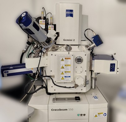
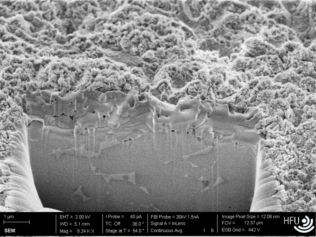
To evaluate the surface quality and microstructural characteristics of the processed samples, scanning electron microscopy (SEM) was employed. The SEM analysis was performed using a ZEISS Crossbeam 550 system, which enabled high-resolution imaging of the nanopore structures and their surrounding areas. As shown in the image, the SEM provided detailed cross-sectional views, allowing precise assessment of surface morphology, layer integrity, and possible material redeposition effects. This characterization was essential for validating the processing parameters and guiding further optimization steps.